
It is humbling to realize that we human share about 70% of our genes with zebrafish. There are also a whole host of other similarities that make these small transparent fish an ideal animal model for the study of many human diseases and biological processes.
In the lab of Dr. Yaniv Elkouby at the Hebrew University of Jerusalem (HU)’s Faculty of Medicine, the focus is on the development of the immature egg cells (oocytes) of zebrafish.
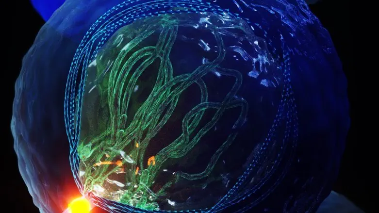
Using unique research tools developed in his lab, researchers were able to watch in real-time as a cluster of oocytes progressed towards maturity. It was during one of these experiments that they saw a hitherto unobserved structure emerging from the cell.
Looking like a twisted fiber, called a cilium, it extended into the mass of surrounding eggs.
Further research showed these cilia play an essential role in chromosomal organization within oocytes. Moreover, they subsequently identified the same structure within the sperm cells of zebrafish and in mouse oocytes and sperm. Their findings were published today in Science (see below for link).
This has implications for their role within human reproduction. Failure of chromosomal organization within human egg and sperm cells result in miscarriages and infertility. However, the mechanisms controlling these processes are not understood. The discovery of a cilium that plays an essential role in controlling chromosomal organization could provide new insights.
Furthermore, defects in cilia formation and function cause genetic disorders called ciliopathies, where patients suffer from deficient fertility and, in tragic cases, babies and children suffer from severe developmental disorders. These were attributed to the failure of other types of cilium. The newly identified cilium provides an additional explanation for these deficiencies. “Identifying mechanisms moves medical research one step closer to finding solutions,” Elkouby shared.
Elkouby pointed out that to explore the function of these new cilia, his team had to apply and develop new advanced methodologies: "We used a repertory of methods, including advanced quantitative and live microscopy, innovative three-dimensional high-resolution imaging, ovary organ culture, manipulations using laser excision, and genetic analyses of multiple mutants."
In this way, his team was able to identify that the newly identified cilium is connected to a "cable-system" within the cell that organizes the chromosomes by mechanically pulling on them. This process is an essential part determining the formation of a fully functional egg that can give rise to healthy offspring. The external cilium anchors the entire cable-system machinery within the egg enabling the essential precise dynamics of the chromosomes to be achieved.
This ground-breaking research, noted Dr. Elkouby, "was a real team-effort, which was co-led by two talented doctoral students: Avishag Mytlis and Vineet Kumar. We also collaborated with the lab of our partner Dr. Sudipto Roy at the institute of Molecular and Cell Biology, Proteos, Singpore, and with the lab of Dr. Ruxandra Bachmann-Gagescu, University of Zurich."