A rare operation to separate a pair of conjoined twins who were attached at their heads was performed last Thursday at Soroka Medical Center.
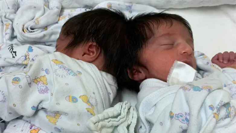
The baby girls were born attached together at the back of their heads in August 2020.
Dozens of staff members from the hospital worked with the family from the moment of birth until the day of the surgery. Preparations were made for their complex birth which included the staff of the Saban Center for Midwifery, doctors and nurses from the Saban Center for Pediatrics, as well as anesthesiologists and imaging specialists.
A dedicated team was gathered for many months ahead of the day of the separation operation.
The separation of Siamese twins who are conjoined at the head is conducted in two steps. In the first stage, the treatment team introduced skin and tissue extenders several months ago in order to produce excess skin that will be used to close the scalp of the two girls after the separation.
Over the past few months the twins have undergone extensive tests, been under continuous monitoring by the Institute for Child Development at Soroka along with the social service that accompanied the family members, in addition to close and close medical monitoring of both of their cardiac and respiratory functions.
They came for regular check-ups at the hospital, and in the last three months have come for more frequent treatment to continue the skin extension operation and prepare for the final stage.
The multidisciplinary team used two models prior to the analysis: 3D models and virtual reality (VR) models. The 3D models of STRATASYS, 3D4OP are based on the images from MRI, CT, and angiography scans and they simulate in the most reliable and accurate way the complexity of the connection of the blood vessels, meninges, skull bones and skin of the twins.
Using the VR model of SURGICAL THEATER, it was possible to make simulations of the procedure and plan it in the most exact manner. Using this model it is possible to go into the depth of the connection between them, see common blood vessels and perform a simulation of all stages of the surgery.
Dozens of repetitions and simulations of all stages of the operation were performed in each of the models with the participation of all the team members.
In the surgery itself, after the separation of the blood vessels was successful, the bones were separated.
At this point, the team members split into two teams that work in parallel in two separate operating rooms, performing a reconstruction of the skull of each of the girls and closing their skin.